Distal Femur Fracture vs. Knee Fracture: Symptoms & Recovery

Chronic Neck Pain from Poor Posture: Lifestyle Modifications & Treatment Options
August 7, 2026
Revision Knee Replacement: Warning Signs, Symptoms & When to Consult an Orthopedic Specialist
August 26, 2026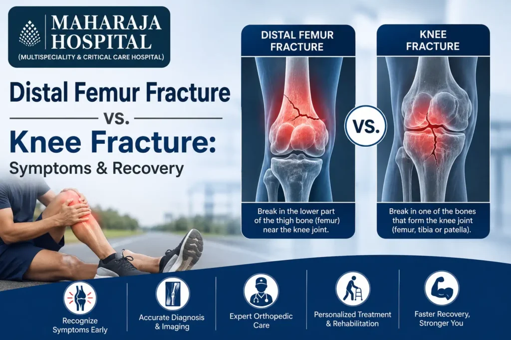
The knee joint is one of the largest, most complex, and most heavily utilized joints in the human body. Because it bears a significant amount of our body weight and facilitates a wide range of motion, it is highly susceptible to injuries, including severe fractures. When a severe impact or a fall occurs, the resulting damage is often broadly referred to as a “broken knee.” However, from a medical and surgical standpoint, fractures in this region are distinctly categorized based on the specific bone involved.
Two of the most critical types of fractures in this area are the distal femur fracture and the general knee fracture (which typically involves the patella or tibial plateau). While they share a geographical location and some overlapping symptoms, their implications for joint mechanics, surgical intervention, and long-term recovery are quite different.
Understanding these differences is vital for patients and their families as they navigate treatment options and physical therapy.
1. What is a Distal Femur Fracture?
The femur, or thigh bone, is the longest and strongest bone in the human body. The “distal” end of the femur is the lower portion that flares out like an inverted funnel and connects with the shin bone (tibia) and kneecap (patella) to form the knee joint. The distal femur is covered in smooth articular cartilage, which allows the joint to glide effortlessly.
A distal femur fracture is a break that occurs exactly in this lower section of the thigh bone. Because the femur is so incredibly strong, it usually takes a massive amount of force to break it in a healthy, young individual.
Common Causes:
- High-Energy Trauma: In younger demographics, these fractures are almost always the result of high-velocity impacts, such as motorcycle accidents, severe car crashes, or falls from a significant height.
- Low-Energy Falls: In elderly populations, particularly those suffering from osteoporosis or weakened bones, a simple fall from a standing position can generate enough force to fracture the distal femur.
- Periprosthetic Fractures: Sometimes, a fracture can occur around a previously installed total knee replacement if the surrounding bone is weak.
2. What Constitutes a General Knee Fracture?
When orthopedic specialists talk about a “knee fracture,” they are generally referring to a break in one of the other two primary bones that make up the knee complex, rather than the thigh bone itself.
Patellar Fracture (Kneecap Break):
The patella acts as a protective shield for the front of the knee joint. It also serves as a fulcrum, increasing the leverage of the quadriceps muscle as it straightens the leg. Fractures here are usually caused by a direct, blunt-force blow to the front of the knee—such as hitting the dashboard in a car collision or falling directly onto a hard surface.
Tibial Plateau Fracture (Shin Bone Break):
The tibial plateau is the flat top surface of the shin bone that supports the distal femur. Fractures here occur when the femur is driven downward into the shin bone. This often happens in falls where the person lands directly on their feet with straight legs, forcing the joint to compress dramatically.
3. Comparing the Symptoms
While both injuries cause immediate, severe pain and an inability to walk normally, there are distinguishing clinical symptoms that help specialists diagnose the exact nature of the injury before taking an X-ray.
| Symptom | Distal Femur Fracture | Knee (Patellar/Tibial) Fracture |
| Location of Pain | Deep ache radiating up into the lower thigh, slightly above the actual knee joint line. | Sharp, localized pain directly on the kneecap or just below the joint line (tibia). |
| Visible Deformity | Leg may appear shortened, crooked, or noticeably bent at an unnatural angle above the knee. | The kneecap may have a visible indentation or gap if shattered; swelling is highly localized. |
| Swelling Location | Massive swelling enveloping the lower thigh and the knee. | Swelling is often contained tightly within the knee capsule itself (effusion). |
| Mobility | Utter inability to bear weight; any attempt to move the leg causes extreme thigh pain. | May be able to put slight weight on it (though not recommended); extreme pain when trying to straighten the leg. |
| Muscle Spasms | Severe quadriceps and hamstring spasms are common as the body tries to stabilize the broken femur. | Spasms are generally less severe, mostly limited to a reflex inhibition of the quadriceps. |
4. Treatment and Surgical Approaches
The treatment path depends entirely on the displacement of the bones (how far out of alignment they are) and whether the fracture breaches the skin (an open fracture).
Treating Distal Femur Fractures
Because the femur bears the entire weight of the upper body, conservative (non-surgical) treatment is exceptionally rare and usually reserved for patients whose medical health prevents surgery. Most distal femur fractures require surgical fixation. Surgeons use plates, screws, or intramedullary nails (a rod inserted down the center of the bone) to hold the pieces together. If the bone is severely shattered, a bone graft might be necessary.
Treating Knee Fractures
- Patellar Fractures: If the kneecap is broken but the pieces haven’t moved out of place, a simple cast or splint keeping the leg straight for several weeks might suffice. If the pieces are pulled apart by the thigh muscles, surgery using pins and tension wires (tension band wiring) is required.
- Tibial Plateau Fractures: Similar to the femur, if the joint surface is depressed or uneven, surgery with plates and screws is mandatory to prevent early-onset arthritis.
When seeking treatment for complex bone injuries, selecting a facility with modern infrastructure is critical. For instance, consulting a highly equipped specialized orthopedic hospital in Rewari ensures that you have access to advanced trauma care, precise imaging, and experienced surgeons who can handle complex joint reconstructions.
Interestingly, the push for minimally invasive techniques has revolutionized all branches of surgery. Just as abdominal issues are now routinely treated with advanced laparoscopic surgeries in Rewari to minimize tissue damage and speed up recovery, orthopedic surgeons also employ minimally invasive plate osteosynthesis (MIPO) and arthroscopic-assisted fracture repairs to achieve similar benefits for joint injuries.
5. Recovery and Rehabilitation Timelines
The recovery journey is often the most challenging part of a fracture. Because both injuries involve a primary weight-bearing joint, rehabilitation is extensive and requires immense patience.
Distal Femur Recovery
The femur takes a long time to heal due to its size and the structural loads it must eventually bear.
- Weight-Bearing: Patients are typically barred from putting any weight on the affected leg for a minimum of 6 to 12 weeks. You will rely entirely on crutches or a wheelchair.
- Motion: Early motion of the knee is encouraged (often using a continuous passive motion machine) to prevent the joint from freezing up.
- Full Recovery: It can take an entire year to regain full strength and a completely normal gait. Some patients may have a permanent slight stiffness in the knee.
Knee Fracture Recovery
- Weight-Bearing: For patellar fractures, weight-bearing in a straight-leg brace can sometimes begin much sooner. Tibial plateau fractures, however, mirror the femur and require weeks of strict non-weight-bearing status to prevent the joint surface from collapsing.
- Physical Therapy: The primary challenge with knee fractures is regaining the final few degrees of extension (straightening) and flexion (bending). Scar tissue builds up rapidly around the kneecap.
- Full Recovery: Generally ranges from 4 to 6 months, though high-impact sports might be off the table for up to a year.
The Role of Physical Therapy
Regardless of whether you break your thigh bone or your shin bone, physical therapy is non-negotiable. The leg muscles—specifically the quadriceps and hamstrings—atrophy rapidly when immobilized. A structured physical therapy program will focus on:
- Passive Range of Motion: The therapist moves the leg for you to keep the joint lubricated.
- Active Range of Motion: You begin moving the leg using your own muscle power.
- Progressive Resistance: Adding weights and resistance bands to rebuild muscle mass.
- Proprioception Training: Balance exercises to retrain the nerves in the leg to understand its position in space.
Conclusion
While both distal femur fractures and general knee fractures result in immediate immobility and significant pain, they are distinctly different injuries requiring specialized care. A distal femur fracture involves the major weight-bearing bone of the thigh and typically demands more invasive surgical repair and a longer timeline before you can walk again. Conversely, knee fractures involving the kneecap or top of the shin bone present their own unique challenges, particularly concerning joint stiffness and arthritis prevention. Prompt medical evaluation, precise surgical intervention, and a grueling but necessary physical therapy regimen are the keys to overcoming either of these devastating injuries.